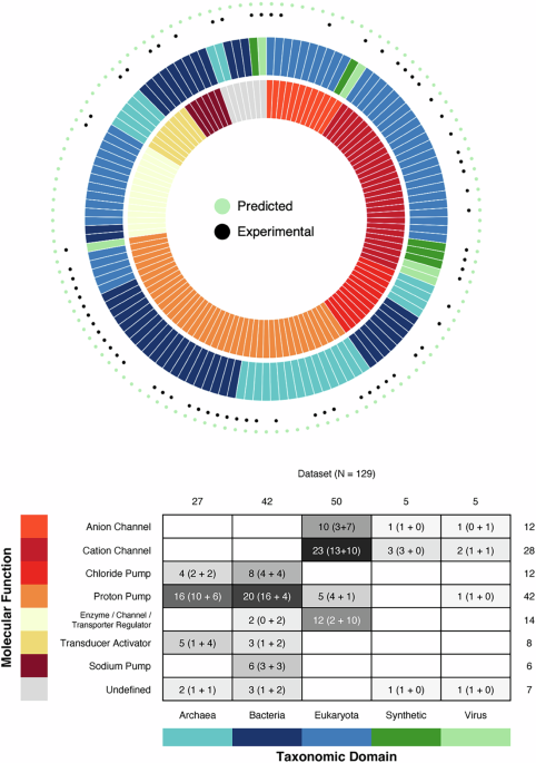
Microbial rhodopsins are photoreceptors with a conserved seven-transmembrane core architecture that use the chromophore retinal—a vitamin A derivative—to convert light absorption into ion transport, signaling, and enzymatic activity. Since their discovery half a century ago, genome sequencing has uncovered thousands of these proteins across archaea, bacteria, eukaryotes, and viruses, revealing a diverse functional repertoire that includes ion pumps and channels, transducer activators, and enzyme/channel regulators. Although the structure of the seven-transmembrane bundle core and the retinal-binding pocket is highly conserved, the sequence identity across major classes is low.
This low sequence identity, combined with the rapidly expanding number of microbial rhodopsins identified by metagenomics, makes direct comparisons across the superfamily increasingly difficult. Here, we introduce a generic residue-numbering system for microbial rhodopsins (MO numbering system), a structure-guided framework anchored to the retinal-binding pocket that enables consistent residue comparison across even the most divergent family members. Analyzing 129 structures and approximately 40,000 sequences, we demonstrate its utility by analyzing sequence–structure–function relationships across the superfamily, including previously uncharacterized members.
The MO numbering system provides a common language for organizing the growing repertoire of microbial rhodopsins, integrating structural, spectroscopic, and electrophysiological data, and engineering next-generation optogenetic tools. The microbial rhodopsin superfamily comprises light-sensitive proteins that use the chromophore retinal—a vitamin A derivative—to convert photon absorption into concerted cellular events. Since the discovery of the proton pump bacteriorhodopsin in Halobacterium salinarum (HsBR) over fifty years ago1, microbial rhodopsins have revealed a striking diversity of functions.
These proteins are found across all domains of life—Archaea, Bacteria, and Eukaryota— and viruses2,3,4, functioning as proton, chloride, or sodium pumps, cation and anion channels, activators of downstream signal transducers, and even as enzyme/channel/transporter regulators. Their photochemical properties are equally varied: while in most microbial rhodopsins light isomerizes all-trans retinal to the 13-cis form, some produce 7-cis, 9-cis, or 11-cis isomers as photoproducts2,5,6,7. Despite this functional and photochemical diversity, all microbial rhodopsins share a common architecture: a tight bundle of seven transmembrane helices (TMs) surrounding the retinal-binding pocket, with retinal bound via a Schiff base to a conserved lysine on TM7.
Some families contain an additional TM preceding TM1, whereas others exhibit an inverted membrane topology6,8,9,10. In addition, individual families adopt characteristic oligomeric assemblies, including dimers, trimers, pentamers, and hexamers6,8,11,12,13,14,15,16. Remarkably, this structural conservation persists even though sequence identity between major families can be as low as 10–12%17, with a nearly identical orientation of retinal in the binding pocket.
Such conservation despite extreme sequence divergence18 suggests powerful evolutionary constraints on the architecture required for retinal binding and photoisomerization. Microbial rhodopsins are often classified, annotated, and compared by tracking characteristic sequence motifs that are coupled to their function. However, the high sequence variability within the family complicates the analysis of large datasets obtained in metagenomics studies, particularly the comparison of distant subfamilies.
Extract — continue reading at the source.


