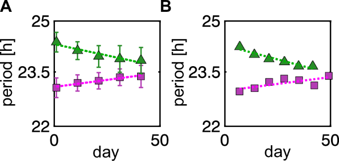
The mammalian circadian pacemaker, the suprachiasmatic nucleus (SCN), comprises a core region that receives light signals from the retina and a shell region that outputs pacemaking rhythms to peripheral tissues. The free-running period (FRP) of animals in constant darkness (DD) correlates with the period of the preceding LD cycle, a phenomenon known as after-effect. To elucidate the mechanisms underlying robust entrainment and after-effect, we analyzed phase oscillator models incorporating attractive and repulsive couplings that respectively decrease or increase phase differences between oscillators.
Attractive coupling from the core to shell regions accounts for experimentally observed phase relationships between these regions under LD cycles. Remarkably, repulsive coupling from the shell to core regions promotes SCN entrainability to LD cycles. Furthermore, after transfer to DD, the FRP slowly returns to the intrinsic SCN period, reflecting the after-effect.
Our analysis identifies an SCN coupling architecture that underlies stable daily activity rhythms. Organisms experience seasonal changes in day–night length throughout the year. Such variations must be internalized by plants1 and animals2 through synchronization of their internal circadian rhythms with external light cycles, a process termed entrainment.
A central question is how the circadian clock achieves stable entrainment to a light–dark (LD) cycle. A classical paradigm for studying this mechanism is to entrain the circadian clock to non-24-h LD cycles referred to as T-cycles3. In mammals, the suprachiasmatic nucleus (SCN) of the brain acts as the central pacemaker of the circadian clock4,5.
Oscillations in the electrophysiological activity of SCN neurons are required to maintain circadian rhythmicity under constant conditions6, and arise from the circadian expression of clock genes7. Among these, delayed negative feedback regulation of Period1/2 (Per1/2) genes is responsible for the generation of autonomous transcriptional rhythms7. The SCN forms a complex network of approximately 104 neurons, divided into ventral “core” and dorsal “shell” regions5,8,9,10.
The ventral region contains neurons secreting vasoactive intestinal peptide (VIP), whereas the dorsal region contains neurons secreting arginine-vasopressin (AVP)8,11. VIP release and AVP production are regulated by neuronal firing activity and circadian clock proteins, respectively12,13,14. These neurons also express both VIP and AVP receptors15,16,17, receiving phase information from neighboring SCN neurons.
Extract — continue reading at the source.


