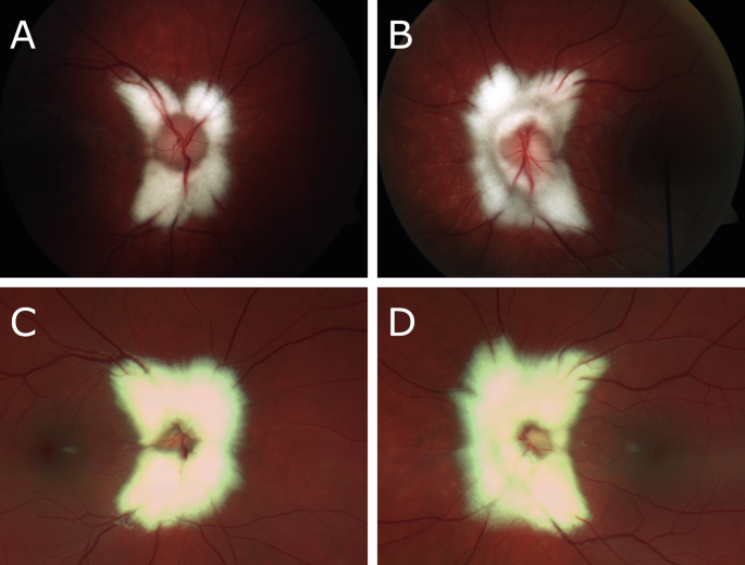
Myelinated retinal nerve fiber layer (RNFL): a comprehensive review. Inter Natl Ophthalmol Clin. 2018;58:147–56. https://doi.org/10.1097/iio.0000000000000239. Rao R, Turkoglu EB, Say EAT, Shields CL.
Clinical features, imaging, and natural history of myelinated retinal nerve fiber layer. Retina. 2019;39:1125–32. https://doi.org/10.1097/iae.0000000000002106. Pan Z, Wei CC, Peng X, Zhang Q, Xu L, Yang H, et al.
Myelinated retinal nerve fiber progression in a 10-year follow-up. The Beijing Eye Study 2001/2011. Am J Ophthalmol. 2021;230:68–74. https://doi.org/10.1016/j.ajo.2021.04.018.
The authors would like to sincerely thank the patient for agreeing to share their ocular imaging findings with the wider medical community. Queen’s University, Kingston Health Sciences Centre, Department of Medicine, Kingston, ON, Canada Metabolism and Systems Science, School of Medical Sciences, University of Birmingham, Birmingham, UK Sean Lazaro: writing the original draft, conceptualisation and resourcing. Daniel Lamoureux: review and editing.
Susan Mollan: conceptualisation, review and editing. SM reports consultancy fees (Novo Nordisk) and advisory board honoraria (SunPharma); Travel (AbbVie, EssilorLuxottica, and European Alliance of Associations for Rheumatology); and receipt of equipment (Heidelberg Engineering). SM is a member of the Eye editorial board.
The other authors declare no potential conflicts of interest. Publisher’s note Springer Nature remains neutral with regard to jurisdictional claims in published maps and institutional affiliations. Open Access This article is licensed under a Creative Commons Attribution 4.0 International License, which permits use, sharing, adaptation, distribution and reproduction in any medium or format, as long as you give appropriate credit to the original author(s) and the source, provide a link to the Creative Commons licence, and indicate if changes were made.
Extract — continue reading at the source.


