Approach–avoidance refers to the psychological conflict in which deciding to approach a reward also carries potential punishment. The orbitofrontal cortex (OFC) is proposed to arbitrate approach–avoidance decisions, yet little is known about the real-time computations within OFC during these decisions. We conducted intracranial recordings in humans implanted with stereotactic electroencephalography electrodes in the OFC while they played a gamified approach–avoidance task.
Here we identified two anatomically distinct signals within the OFC prior to approach: an increase in activity within the medial compartment and a decrease within the lateral compartment. Prior to decisions, these signals rapidly alternated between discrete pro-approach and pro-avoidant states, thus introducing a new functional architecture within the human OFC during decision-making. Approach–avoidance conflict arises when an action carries potential rewards and punishments, leading to decision conflict1.
In nonhuman primates (NHPs), rewarding and punishing stimuli across sensory modalities converge in the orbitofrontal cortex (OFC)2, and many OFC neurons signal economic value3,4,5,6, making the OFC an obvious candidate for shaping the decision to approach or avoid1. There is a paucity of studies that delve into the functional and anatomical underpinnings of approach–avoidance decision-making in human OFC. The human OFC sits atop bone and near air-filled sinuses, constraining functional magnetic resonance imaging (MRI) findings7.
Furthermore, increased reaction time, a hallmark of decision conflict, increases functional MRI blood oxygen level-dependent signals8. Approach–avoidance studies controlling for reaction time yielded mixed results on the representation of decision-related variables in the OFC9,10, underscoring the need for higher temporal resolution methods to capture the computations underlying approach–avoidance decisions. We harnessed intracranial electrode recordings in patients implanted for clinical indications.
In keeping with standard clinical care, OFC electrodes were implanted medial to lateral11, allowing us to simultaneously capture activity across OFC subregions. An NHP study demonstrated that high-frequency activity (HFA; 70–150 Hz), which indexes firing of neural ensembles, captures OFC value coding similar to single-unit recordings12. By analyzing HFA along the horizontal anatomical axis in humans, we recorded a wide swath of OFC anatomy with excellent temporal resolution.
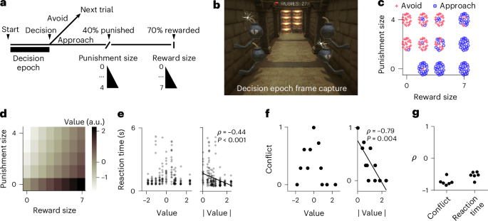
Six patients (Extended Data Table 1) completed an approach–avoidance task with the goal of earning rubies (Fig. 1a,b). On each trial, the participants viewed a corridor containing bombs and treasure chests (Fig. 1b). Following a 600-ms pan-in period before responses were allowed, the participants had 6 s (‘decision epoch’) to decide whether to approach or avoid.
Extract — continue reading at the source.


