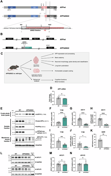
While the accumulation of Aβ peptides, derived by proteolytic cleavage from the amyloid precursor protein APP, constitutes a hallmark of Alzheimer’s disease, the question of whether endogenous, physiological APP processing serves an essential role for normal brain function(s) has remained elusive. Here, we modified the mouse APP locus to delete the endogenous α- and β-secretase sites (APP residues S592-G621) while preserving the transmembrane topology and the APP C-terminus. These novel knockin mice (termed APPΔS622) lack APPsα, APPβ, and Aβ production and show only minimal soluble APPs shedding.
These APPΔS622 mice provide a unique model to distinguish between functions mediated by unprocessed cell surface APP involved in synaptic adhesion or by proteolytic fragments. Strikingly, APPΔS622 mice exhibit pronounced impairments in both long-term potentiation (LTP) and homeostatic forms of synaptic plasticity. Extracellular field recordings revealed significant deficits in hippocampal LTP, associated with a reduced density of mature spines and reduced spine head size.
LTP deficits could be restored by APPsα application, suggesting a requirement for non-amyloidogenic APP processing for LTP. In addition, APPΔS622 mice lacking endogenous Aβ displayed a lack of homeostatic synaptic scaling in response to network impairment that could be specifically rescued by exogenous Aβ. Importantly, these deficits in different forms of synaptic plasticity also led to impairments in cognitive behavior.
In summary, our findings reveal an essential role of physiological APP processing products to differentially regulate complementary forms of synaptic plasticity that are critical for network stability, normal brain function and cognition. Despite the molecular cloning of APP more than 30 years ago [1,2,3] and the subsequent genetic linkage of autosomal dominant APP mutations to familial forms of Alzheimer’s disease (AD) [4], the normal, physiological role of APP in the brain remains poorly understood. Strikingly, it is still unclear whether APP processing as such has an essential physiological role for brain function.
In AD, synaptic dysfunction and loss of synapses occur early in the course of the disease, prior to overt Aβ accumulation. Importantly, these early synaptic aberrations are highly correlated with disease severity and cognitive decline [5,6,7]. Although recently developed Aβ-directed immunotherapies can somewhat slow down cognitive decline, clinical benefit is small and associated with vascular side effects including hemorrhage [8, 9].
To improve therapeutic strategies, it is therefore pivotal to better understand the physiological role of APP for synapse formation, function, and plasticity in health and disease. APP is a type I transmembrane protein that is highly expressed across cell types in the brain, with particularly high expression during synaptogenesis. It is present at both pre- and postsynaptic sites and constitutes approximately 1% of all synaptic proteins [10,11,12].
Extract — continue reading at the source.


