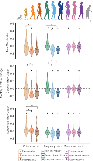
The female brain undergoes substantial reorganization during major hormonal transitions, yet whether puberty, pregnancy, and menopause engage shared or distinct neuroplastic mechanisms remains unknown. We compared longitudinal structural brain changes across all three major hormonal life events using identical analytical methods within a single cohort framework (n = 1095), with stable control groups to control for normative developmental and aging trajectories. Both pre-to-post menarche and pre-to-post pregnancy groups showed widespread cortical gray matter reductions, with cross-cohort comparisons revealing divergent profiles across more than half of cortical regions alongside convergence in prefrontal, parietal, and temporal association cortices.
Pre-to-post menopausal women showed a fundamental divergence from both puberty and pregnancy, as they showed no volumetric loss, while stable pre- and postmenopausal control groups did. Puberty and pregnancy thus share both convergent and divergent cortical remodeling, while menopause represents a qualitatively distinct pattern, defined by the absence rather than acceleration of volumetric change. The female brain can undergo three major hormonal life events across the lifespan: puberty, pregnancy, and menopause.
Each event involves profound shifts in sex steroid hormones, particularly estrogens and progesterone, which bind to widespread receptors throughout the brain. Longitudinal neuroimaging studies have begun to characterize structural brain changes during puberty and pregnancy, but evidence for menopause is limited to a single study that did not examine whole-brain gray matter structure. Whether these three events engage a shared neuroplastic program or each triggers distinct patterns of remodeling have not yet been directly examined.
Puberty represents the first major hormonal life event, characterized by dramatic increases in sex steroid hormones that coincide with extensive brain remodeling. Although most studies focus on the entire adolescence period, emerging research has begun to isolate puberty-specific effects on brain structure. Longitudinal neuroimaging studies document reductions in cortical gray matter volume across puberty1,2,3,4,5,6,7.
Recent evidence suggests that menarche, the first menstruation, may mark a particularly salient transition point within this broader developmental window, as machine learning algorithms can classify pre- versus post menarche status from brain structure with high accuracy8. These findings suggest that the menarche transition itself represents a discrete neuroplastic event warranting specific investigation, rather than simply one point along a continuous pubertal trajectory. Similarly, pregnancy induces pronounced structural brain changes.
Longitudinal studies comparing pre-conception to pregnancy and post-pregnancy brain scans in women becoming first-time mothers by our and other groups reveal decreases in gray matter volume9,10,11,12,13,14. Reductions are linked to estradiol levels during pregnancy10, and some persist for at least six years postpartum15. Recent work from our group shows similar changes occur during a second pregnancy, with some specific differences11.
Extract — continue reading at the source.


