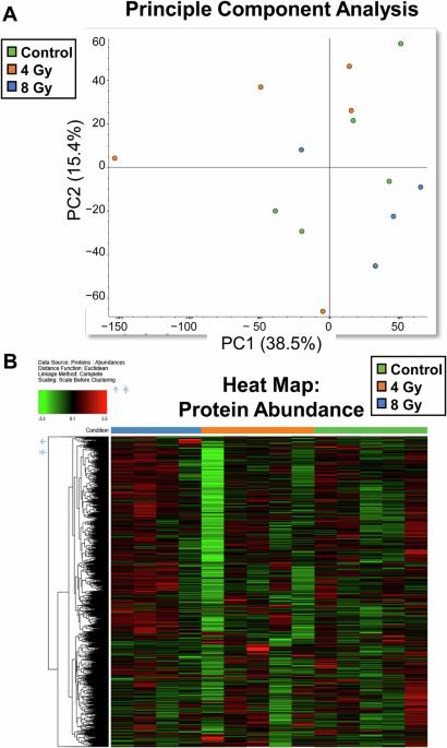
Long-term neurological consequences of acute total-body γ-radiation are poorly understood. Here we investigated the persistency of molecular changes in non-human primates (NHPs) 5+ years after single exposure to ~4 Gy or ~8 Gy total body irradiation dose. MS-based proteomic analyses were performed on samples of micro-dissected hippocampus from brains of irradiated/control NHPs.
We used STRING and DAVID analyses to expand interpretation of proteomic data. Proteomic findings were further assessed using Western blot and digital-PCR. Proteomic analysis of the hippocampus revealed profound, dose-dependent molecular changes. ~4 Gy primarily impacted synaptic plasticity, while ~8 Gy affected catabolism, vascular development, and inflammation.
Apelin receptor (APLNR) was the only protein consistently lowered at both radiation doses, suggesting it may be a key biomarker for radiation-induced brain injury. Significantly, while most proteins linked to neurodegeneration were unchanged, Tau (MAPT) protein levels increased at ~8 Gy, and some animals developed Tau-positive lesions resembling neurofibrillary tangles. Moreover, we found complex, dose-specific alterations in phosphorylated forms of other key proteins (alpha-synuclein, TDP-43).
Overall, these findings show that different radiation doses induce distinct, persistent molecular changes in NHP hippocampus. This work highlights dysregulation of proteins usually associated with neurodegeneration (i.e., Tau and synaptic proteins) as a major long-term consequence of radiation, providing new insights into the molecular shift that accompany post-exposure high-dose radiation and likely a possible cause of post-exposure cognitive deficits. Exposure to high-dose γ-radiation can cause severe and life-threatening health consequences, including acute radiation syndrome (ARS), late delayed radiation-induced brain injury (RIBI), and the delayed effects of acute radiation exposure (DEARE), which often manifest as chronic, multi-organ damage1,2,3.
To better understand and mitigate these risks, particularly following accidental or intentional radiation events, the use of a translationally relevant animal model is essential. Non-human primates (NHPs) are considered the gold standard for such studies due to their close physiological, immunological, behavioral, and genetic similarities to humans, which allow for more accurate investigations of radiation-induced pathology, and brain pathology in particular1,4. Among the organ systems vulnerable to radiation, the central nervous system (CNS) is of particular concern due to its integral role in an organism’s overall health and function.
However, the long-term effects, their molecular mechanisms, and late clinical manifestations of sublethal γ-radiation exposures to the brain tissue and function remain poorly understood. Previous studies in swine models have provided some initial insight into the effects of a single acute total-body low-dose (1.79 Gy) γ-radiation exposure on various molecules and anatomical regions of the brain5. These recent findings in swine revealed that an acute total-body low-dose sublethal radiation exposure can induce complex region-specific and molecular alterations across various regions of the brain, which may be either detrimental or beneficial if related to a specific clinical or occupational context6,7.
Extract — continue reading at the source.