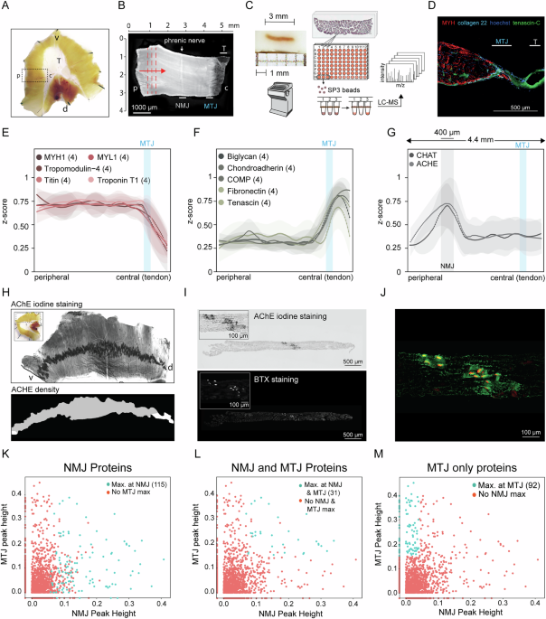
Morphological studies of the diaphragm have provided a detailed view of its architecture, which consists of parallel skeletal muscle fibers and a central ring of neuronal innervation that includes neuromuscular junction (NMJ) units. NMJs are disease-vulnerable synapses; thus, analysis of the NMJ is essential to understand its function in both healthy and disease-related conditions. The diaphragm was analyzed by spatial proteomics and 115 proteins were enriched at the NMJ.
Comparison of the protein signatures of the NMJ and myotendinous junction (MTJ) revealed 31 shared proteins, suggesting partially conserved structures between these junctions. Key mediators of synaptic transmission and extracellular matrix organization were observed among the NMJ-enriched components, which indicates the molecular complexity and regulatory potential of the NMJ. A focused study of the uncharacterized NMJ protein FXYD6 demonstrate enhanced FXYD6 expression in type IIa fibers of the diaphragm, which exhibit a unique balance of oxidative and glycolytic capacity.
FXYD6 interacts with Na⁺/K⁺-ATPase subunits in the diaphragm, which supports the function of FXYD6 in the ionic homeostasis required for continuous, fatigue-resistant contraction. Overall, the dataset provides a comprehensive molecular atlas of the NMJ in the diaphragm and opens new opportunities to dissect the synaptic mechanisms underlying respiratory function and neuromuscular diseases. The diaphragm is a constantly contracting muscle that is innervated by the bilateral phrenic nerves, and neuromuscular junctions (NMJs) convert motor neuron action potentials into chemical signals that trigger post-synaptic depolarization and muscle contraction1,2.
In addition, motor neurons instruct the myonuclei at the synapses to express specific genes, and locally regulated protein synthesis may coordinate the assembly of these functional compartments within the muscle. However, a comprehensive proteomic analysis of NMJ structures is essential to elucidate the precise molecular composition of their pre- and post-synaptic components, including the neighboring cells and extracellular matrix (ECM) proteins. This knowledge will provide mechanistic insights into how NMJs regulate synaptic activity, maintain structural integrity, and control skeletal muscle contraction.
Synaptic transmission at the NMJ starts when an action potential reaches the presynaptic terminal of a motor neuron, which causes the neurotransmitter acetylcholine (ACh) to be released and diffuse across the synaptic cleft. ACh then binds to nicotinic acetylcholine receptors (nAChRs) on the cell membrane of muscle fibers to induce a post-synaptic action potential. This activation triggers sarcolemmal release of calcium, which binds to troponin C and induces skeletal muscle contraction3.
Sodium and potassium ATPases (Na⁺/K⁺-ATPase) play crucial roles in maintaining the ion gradients necessary for action potential generation and propagation. These ATPases actively transport sodium ions from outside the plasma membrane into the muscle fiber to restore resting membrane potential after depolarization. The motor endplates of NMJs form specialized nerve-muscle contact sites.
Extract — continue reading at the source.


