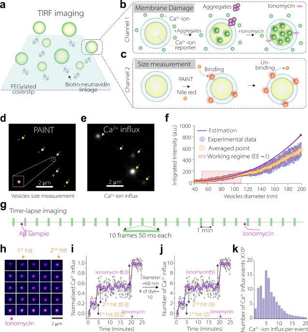
Neurotoxic intermediates formed during amyloid-β (Aβ) aggregation are thought to drive Alzheimer’s disease, yet their rarity and transient nature have obscured their identity and formation pathways in the human brain. Here, we combine super-resolution microscopy and single-molecule functional imaging with kinetic modelling to classify Aβ intermediates according to their membrane-disrupting activity and connect their damaging effects to the aggregation pathways that generate them. Applying this framework to recombinant Aβ42, an Alzheimer’s knock-in mouse model and human Alzheimer’s brain tissue, we find two functionally distinct intermediate populations present across all systems.
They form through separate aggregation routes and differ in their neurotoxicity, abundance and susceptibility to inhibition. Both damage neurons and glia, but only one converts into plaques. Suppressing these intermediates pharmacologically in mice reduces plaque burden, attenuates astrogliosis and improves cognition.
Our findings connect model systems with human pathology and identify intermediate formation pathways as actionable therapeutic targets across protein-misfolding disorders. Alzheimer’s disease (AD) is the most prevalent form of dementia, affecting over fifty million individuals worldwide. One of its defining pathological hallmarks is the extracellular deposition of amyloid-β (Aβ) peptides into plaques1, a process that begins with the misfolding and aggregation of Aβ monomers into soluble intermediates, which subsequently assemble into insoluble fibrils that accumulate as amyloid plaques2,3.
As Aβ aggregates and deposits in the brain, levels of soluble Aβ in cerebrospinal fluid decline, providing one of the earliest detectable abnormalities in AD4. Yet the relevance of this aggregation pathway to disease progression remains contentious: Aβ plaque burden correlates poorly with cognitive decline5,6, and immunotherapies aimed at clearance of plaques have shown only modest clinical benefit7,8. These observations raise fundamental questions about the causal role of Aβ plaques in AD9,10.
Mounting evidence instead implicates Aβ intermediates, such as oligomers, formed en route to plaques, as key drivers of neurodegeneration and better correlate with cognitive dysfunction11,12,13,14,15,16. Among the Aβ isoforms, the 42-residue Aβ peptide, Aβ42, is particularly prone to aggregation17 and disproportionately enriched in amyloid plaques in AD brains18. It is also deposited into amyloid plaques earlier in the disease process compared to other Aβ isoforms19.
As a result, the role of Aβ42 in AD is extensively studied in model systems, primarily through recombinant protein assays and transgenic animal models. Whilst these approaches have provided key insights into Aβ dysfunction and associated toxicity, they have certain limitations. Recombinant studies typically require Aβ concentrations several orders of magnitude higher than those found in the human brain20.
Extract — continue reading at the source.


