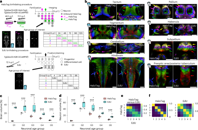
Understanding how neurons integrate into developing circuits and contribute to functional activity is essential for decoding brain development and plasticity. However, current methods to study neuronal integration often suffer from low throughput, limited spatiotemporal resolution or invasive procedures. To overcome these challenges, here we present CLOK, an in vivo birthdate-labeling strategy based on HaloTag technology and its broad palette of fluorescent ligands.
We show that CLOK enables precise color-coding of neurons and their subcellular structures by birthdate and allows tracking of functional maturation of newborn neurons in the developing zebrafish visual and motor systems, revealing distinct maturation trajectories of early- and late-born neurons. We further demonstrate the versatility of this approach by coupling it to optical tools enabling age-specific multicolor calcium and voltage imaging, as well as optogenetic manipulation. Altogether, CLOK provides a powerful and flexible tool for optical interrogation of neuronal circuit maturation in a growing and behaving vertebrate.
Understanding the developmental trajectory of individual neurons and the maturation of their roles within neural circuits is essential to elucidate brain development and plasticity during both embryonic and postembryonic growth, when progressively more refined functions emerge. The integration of neurons into developing circuits has been extensively investigated across vertebrate models1,2,3. Newly generated neurons are gradually incorporated into existing circuits by extending axons and dendrites and forming synaptic connections4.
In several regions of the central nervous system, neurogenesis is spatially organized, with newborn neurons being progressively added in topologically distinct areas. Examples include the spinal cord5, hippocampus6, cortex and cerebellum7. In this context, the zebrafish larva offers a particularly advantageous model for studying neurogenesis and functional integration owing to its transparency and rapid development3,8.
Importantly, a similar topographical organization is observed in the zebrafish nervous system, where some regions exhibit a highly ordered structure characterized by a developmental gradient9,10,11. A striking example is the optic tectum (OT), the main visual center in the fish brain: neurons generated early in development (early-born) occupy central regions, while newly generated neurons (late-born) are progressively added to peripheral regions throughout the animal’s life12,13. Previous studies were limited by low throughput and methodological constraints, underscoring the need for more advanced tools to examine neurogenesis and functional integration at the population level, ideally in vivo.
Indeed, a comprehensive functional and causal study of neurogenesis and neuronal integration requires a method capable of identifying the maturation stage of individual neurons and distinguishing early-born from late-born neurons within the intact, developing brain. Furthermore, such an approach should also enable the monitoring of neuronal activity patterns, particularly in relation to specific behaviors or sensory inputs, and permit noninvasive manipulation of neuronal activity. This capability would facilitate precise correlations between neuronal activation, downstream circuit dynamics and behavioral outcomes.
Extract — continue reading at the source.