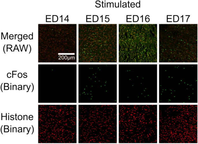
The timing of functional hypothalamic responsiveness during embryogenesis remains incompletely defined. We examined hypothalamic activation in chicken embryos from embryonic day (ED) 12–17 following experimental stimulation consisting of a localized foot injection of paraformaldehyde (PFA) together with the associated handling procedures required for its administration. Hypothalamic activation was quantified as the proportion of c-Fos-positive cells within the paraventricular nucleus (PVN).
Generalized linear modelling revealed significant effects of both embryonic age and stimulation condition on c-Fos expression, with no evidence of an Age × Stimulation interaction. Across development, stimulated embryos exhibited higher proportions of c-Fos-positive cells than age-matched controls. Independent of stimulation, the only significant difference between adjacent developmental stages occurred between ED14 and ED15, suggesting a candidate transition in hypothalamic maturation.
Exploratory analysis indicated that these findings are consistent with an early (ED12–ED14) and a late (ED15–ED17) developmental phase and suggest that ED14–ED15 may represent a candidate phase boundary in hypothalamic maturation. The functional maturation of neural circuits during embryonic development is a gradual and highly coordinated process1,2. Although structural development of major brain regions has been extensively characterized across vertebrate species, the timing at which these structures acquire functional responsiveness to sensory input remains incompletely defined3,4.
In particular, the developmental onset of central responsiveness to noxious stimulation has attracted increasing attention, as it provides insight into the maturation of sensory integration and stress-regulatory systems5,6. Nociceptive processing develops hierarchically, involving peripheral sensory neurons, ascending brainstem pathways, subcortical integrative centers, and ultimately higher-order cerebral circuits7,8,9,10. Experimental studies across species indicate that early responses to noxious input are mediated primarily by subcortical and brainstem structures, with cortical integration emerging later in development5.
Functional activation markers such as c-Fos are widely used to map stimulus-induced neuronal recruitment and to delineate the maturation of central circuits11,12. In rodents, stimulus-induced Fos expression in central nociceptive pathways increases postnatally, reflecting progressive circuit maturation6. These findings suggest that the acquisition of central responsiveness is not instantaneous but instead follows distinct developmental stages.
Among subcortical structures, the hypothalamus occupies a pivotal position in coordinating autonomic and neuroendocrine responses to stress-related and sensory stimuli13. The paraventricular nucleus (PVN) integrates ascending sensory input with endocrine and autonomic outputs and is a key regulator of hypothalamic–pituitary–adrenal (HPA) axis activation14,15. While the structural development of hypothalamic nuclei has been described in multiple species16, considerably less is known about the developmental timing of functional hypothalamic activation in response to peripheral noxious stimulation.
Extract — continue reading at the source.


