The human brain undergoes profound changes from early development through late adulthood, shaping cognition, behaviour and vulnerability to disease1,2. Understanding how these changes are organized within specific brain regions and cell types is essential for interpreting normal ageing and its relationship to psychiatric and neurodegenerative disorders. The dorsolateral prefrontal cortex has a central role in higher cognitive functions and is particularly sensitive to age-related decline3, yet its cellular and molecular programs across the human lifespan remain poorly defined.
Most existing studies4,5,6,7 have focused on restricted age ranges or disease-affected brains, limiting the ability to distinguish normative developmental and ageing trajectories from pathological processes. Consequently, a comprehensive, lifespan-resolved reference of cellular states in the human prefrontal cortex has been lacking. Here, using a single-nucleus transcriptomic atlas spanning the human lifespan, we show that the dorsolateral prefrontal cortex exhibits non-linear, cell-type-specific transcriptional trajectories characterized by dynamic remodelling during development, relative stability in midlife and selective molecular reactivation in late adulthood.
We identify distinct neuronal and glial programs, including early-life neuronal resilience pathways and late-life glial programs associated with immune activation, stress responses and circadian reorganization. These programs are anatomically organized across cortical layers and grey–white matter domains, revealing coordinated spatial and molecular changes. Together, these findings provide a framework for understanding how cellular programs transition from resilience to vulnerability in the human cortex and establish a foundation for interpreting age-related cognitive decline and disease risk.
The organization of molecular and cellular programs across the human lifespan remains a central challenge in understanding brain development, ageing and disease susceptibility. Although single-cell transcriptomic studies4,5,6,7 have transformed our understanding of cellular diversity in the human brain, how transcriptional programs evolve continuously from early development through late adulthood within defined cortical regions remains poorly characterized. The dorsolateral prefrontal cortex (DLPFC), which supports higher cognitive functions3, is particularly well suited for investigating lifespan dynamics given its prolonged maturation and sensitivity to age-related decline.
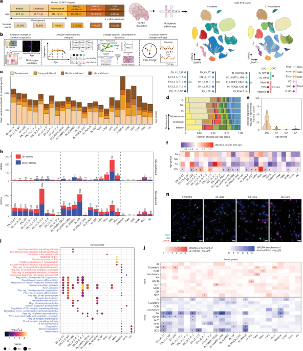
To address this gap, we generated a single-nucleus transcriptomic atlas of DLPFC spanning the full lifespan, profiling over 1.3 million nuclei from neurotypical donors. This resource, produced as part of the PsychAD Consortium8,9, enables systematic analysis of cell-type-specific transcriptional programs across development, adulthood and ageing. Using integrated differential expression, trajectory modelling and spatial transcriptomic analyses, we identify three major phases of molecular change, define age-dependent gene modules within neuronal and glial lineages, and link these programs to genetic risk for psychiatric and neurodegenerative disorders.
We further identify age-related reorganization of circadian gene expression, marked by loss of neuronal clock synchronization and the emergence of glial stress-associated rhythms. Together, these findings establish a framework for understanding how cellular programs transition from resilience to vulnerability across the human lifespan. To construct a lifespan-resolved transcriptomic atlas of the DLPFC at the single-nucleus resolution, we profiled single-nucleus RNA-sequencing (snRNA-seq) libraries from 284 neurotypical donors spanning infancy (0–1 years old), childhood (2–11 years old), adolescence (12–19 years old), and young (20–39 years old), middle (40–59 years old) and late (≥60 years old) adulthood8,9 (Fig. 1a, Supplementary Fig. 1a, Supplementary Table 1, Supplementary Data 1 and Supplementary Notes).
Extract — continue reading at the source.