Tuberous sclerosis complex (TSC) is a genetic neurodevelopmental disorder characterized by focal brain malformations called cortical tubers, which are associated with severe, intractable epilepsy1. Tubers are thought to result from somatic second-hit mutations that disrupt the TSC1 or TSC2 genes in neural progenitor cells, driving hyperactive mTORC1 signalling2. Glial abnormalities are commonly observed in tubers3; however, whether glia actively cause disease or merely result from chronic seizure activity has remained unclear.
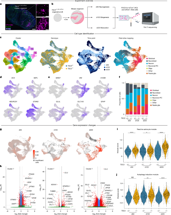
To address this question, we used human brain organoid models to track the developmental fate of mutated progenitor cells in the absence of seizures. Here we show, using single-cell transcriptomics and cyclic immunostaining across human brain organoids and resected tuber tissue from patients, that loss of TSC2 biases neural progenitors to differentiate into enlarged, pro-inflammatory reactive astrocytes in a cell autonomous manner. These mutant astrocytes show downregulated glutamate transporter expression, increased inflammatory cytokine secretion and elevated expression of neurodegenerative disease risk genes such as APOE and CLU.
Our findings demonstrate that reactive astrocytes emerge as a primary consequence of TSC2 loss. These results implicate glial dysfunction as a driver of TSC pathogenesis and highlight reactive astrocytes as potential therapeutic targets for TSC-related neuropathology. Tuberous sclerosis complex (TSC) is a multisystem developmental disorder caused by mutations in TSC1 or TSC24.
TSC leads to hamartomas in several organs and a constellation of neurological and psychiatric conditions, including severe, early-onset epilepsy1. Malformations of cortical development, known as cortical tubers, are a hallmark of TSC5. A subset of tubers and surrounding regions can become seizure foci, and surgical removal can be beneficial for individuals with intractable epilepsy6.
The origin of tuber cells and the molecular changes that lead to their abnormal development are open questions. On a cellular level, tubers contain heterogeneous populations of dysmorphic neurons, dysplastic and gliotic astrocytes, activated microglia and giant or balloon cells within a background of normal-appearing cells7,8,9. Although much research has focused on neuronal mechanisms in TSC10,11,12,13, some studies have suggested that the primary pathological cells within tubers may be astrocytes3.
Indeed, gene-expression analysis of tuber tissue has revealed increased expression of astrocyte-enriched genes and prominent neuroinflammation14,15. However, because tuber resections are performed in individuals with intractable epilepsy, it has been unclear whether glial changes are a cause or consequence of continuing seizures. A further challenge is the lack of a clear cell type signature within tuber lesions, as giant cells can express progenitor cell markers as well as neuronal and glial proteins8,16,17.
Extract — continue reading at the source.