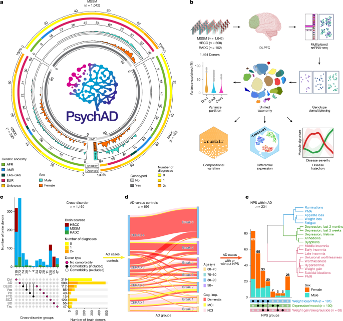
Neurodegenerative and neuropsychiatric diseases impose a considerable societal and public health burden. However, our understanding of the molecular mechanisms underlying these highly complex conditions remains limited1,2. Here, to gain deeper insights into the aetiology of different brain diseases, we used specimens from 1,494 unique donors to generate a population-scale single-cell transcriptomic atlas of the human dorsolateral prefrontal cortex, comprising over 6.3 million individual nuclei.
The cohort includes neurotypical controls, as well as donors affected by eight common and complex brain disorders: Alzheimer’s disease (AD), diffuse Lewy body disease (DLBD), vascular dementia (Vas), Parkinson’s disease (PD), tauopathy, frontotemporal dementia, schizophrenia, and bipolar disorder. We show that interindividual variation accounts for a substantial portion of gene expression variation. By comparing transcriptomic variation across diseases, we reveal universal signatures enriched in basic cellular functions such as mRNA processing and protein localization.
After discounting these cross-disease signatures, we show stronger genetic and transcriptomic concordance among AD, DLBD, Vas and PD. Furthermore, we characterize transcriptomic variation among different AD phenotypes, distinct from those observed in healthy ageing, revealing a reduction in neuronal abundance in individuals with more severe AD, coupled with an increase in immune and vascular cell populations. Exploring the neuropsychiatric symptoms (NPSs) that frequently accompany AD, we find an increased abundance of deep-layer excitatory neurons associated with a broad range of NPSs.
By constructing transcriptome trajectories that capture AD progression, we implicate cell-type-specific responses in the early and late stages of AD. Our disease atlas provides a perspective of the transcriptomic landscape in neurodegenerative and neuropsychiatric disorders, shedding light on shared and distinct processes involving the neurological–immune–vascular systems, and identifying potential targets for therapeutic intervention. The human brain is a highly complex organ composed of billions of functionally diverse cells.
Under pathogenic stress, cellular and molecular responses are often convoluted and contextual, so understanding their dysfunction in disease is challenging. Single-cell approaches that facilitate analysis of the molecular changes that occur within individual cells have been particularly helpful in understanding the interplay between the different cell types found within complex tissues, as well as the roles of those cells in various disease contexts. In AD, a thorough exposition of cellular heterogeneity in the brain has revealed that studying the coordinated interactions between neurons and glia, and the selective depletion of vulnerable inhibitory neuronal subtypes, is critical for understanding AD pathology1,2.
Building a large-scale disease atlas at the single-cell resolution creates an opportunity to understand molecular responses at the cellular level and estimate population-level variation in the brain transcriptome. A large sample size provides the power needed to establish robust basal-level conditions and to sufficiently capture the full spectrum of disease pathology. By leveraging cross-disease atlases, studies have revealed shared and distinct patterns of gene expression perturbations in major psychiatric diseases, as well as shared genetic factors leading to molecular convergence3,4.
Extract — continue reading at the source.


