Alzheimer disease begins in the brain many years before symptoms, but early changes are usually studied after death or indirectly through fluid and imaging biomarkers. Here we show that small brain biopsies collected during ventriculoperitoneal shunt surgery can be used to study Alzheimer disease pathology and protein turnover during life. We analysed biopsies from 18 individuals with suspected normal pressure hydrocephalus, alongside matched ventricular and lumbar cerebrospinal fluid and post-mortem control brain tissue.
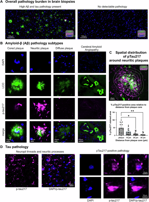
Amyloid plaques were present in 9 of 18 biopsies, while mature tau tangles were rare. Matrix-assisted laser desorption/ionization mass spectrometry imaging mapped amyloid plaque chemistry within tissue. Phosphorylated tau 217 was enriched around plaques, supporting a local relationship between amyloid and tau pathology.
Stable isotope labelling, which tracks newly made proteins, detected rapid tau labelling within approximately 3 hours and estimated brain tau half-life at about 34 days. Amyloid plaques showed little detectable turnover. Alzheimer disease (AD), the commonest cause of dementia, is characterised by deposition of β-amyloid (Aβ) peptide derived from amyloid precursor protein (APP) and intracellular deposition of hyperphosphorylated tau protein1,2,3.
These neuropathological observations are based on post-mortem examination of the major pathological hallmarks, i.e. extracellular Aβ plaques; extracellular neuritic plaques; intracellular tau tangles4,5 and supported by cerebrospinal fluid (CSF), plasma6,7,8 and imaging biomarkers (Aβ and tau Positron Emission Tomography (PET))9,10,11,12,13,14,15. We lack understanding of the evolution of Aβ and tau pathologies in human brain tissue, particularly at earlier stages of the AD continuum. This is due to limited access to human brain tissue during life, and limitations in plasma and CSF biomarkers for spatial and temporal biochemical characterization of earliest neuropathologic AD changes in the brain16,17.
This is especially important as recent advancements in fluid biomarkers implicate different Aβ and phosphorylated tau (pTau) species at earlier stages of the AD continuum18,19,20,21. Until recently, technical limitations have prevented detailed biochemical characterization of Aβ and tau pathological structures at cellular and sub-cellular level, with limited means of determining the rate at which proteins are synthesized, accumulate, are cleared or modified over time22,23,24. Understanding how these structures form, how they evolve over time and how Aβ and tau interact ex vivo has implications for therapeutic intervention, particularly at the earliest pre-symptomatic phase of the disease25,26,27,28.
A number of complementary techniques can now help to overcome these problems. Immunoprecipitation (IP) combined with targeted mass spectrometry (MS) can provide high resolution and detailed information on the biochemical changes in the protein when soluble and insoluble protein fractions are obtained. However, it has its own challenges such as poor spatial visualisation29.
Extract — continue reading at the source.


