Metabotropic glutamate receptors (mGluRs) are prototypical, dimeric family C G protein-coupled receptors (GPCR) that perform crucial modulatory roles throughout the nervous system. While mGluR activation and signaling through G proteins has been studied extensively, how these receptors interact with and are desensitized by β-arrestins (β-arrs) is not well understood. Here, we use an integrative biophysical and structural approach to probe the coupling of mGluR8 and β-arrs.
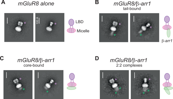
Using negative stain electron microscopy (EM), we identify tail- and core-bound orientations and stoichiometries of mGluR8/β-arr complexes. Cryo-EM structures of mGluR8 alone or bound to either G proteins or β-arr1 reveal mGluR8 active states with transducer-specific differences. The mGluR8/β-arr structure shows a distinct complex orientation compared to other GPCR/β-arr structures which supports a steric mechanism of mGluR desensitization involving interactions with both subunits and the lipid bilayer.
Coupling of mGluR8 to β-arr1 in an active-like conformation is verified by live-cell and single molecule FRET analysis. Finally, molecular dynamics simulations further define the positioning and dynamics of mGluR8-bound β-arr1 and the importance of critical mGluR8 residues for stabilizing β-arr1 complexes. Together, our data provide a framework for agonist-driven family C GPCR/β-arr coupling.
Metabotropic glutamate receptors (mGluR) are family C G protein-coupled receptors (GPCRs) that play central roles in various modes of neuromodulation throughout the central and peripheral nervous systems1,2. Generally, mGluRs are categorized into three groups based on their sequence identity, G protein-coupling properties, and synaptic localization. Gq-coupled group I mGluRs (mGluR1, mGluR5) and Gi/o-coupled group II mGluRs (mGluR2, mGluR3) represent the highly studied, prototypical post- and pre-synaptic mGluR subtypes, respectively.
The group III mGluRs (mGluR4, mGluR6, mGluR7, mGluR8) are also Gi/o-coupled and primarily pre-synaptic, but less well studied than their group I and group II counterparts. However, group III mGluRs represent promising drug targets for a variety of neurological and neuropsychiatric disorders. Their therapeutic potential is underscored by their restricted expression profiles and lower expression levels compared to group I/II mGluRs, which may provide a basis for safer, more modulatory targeting.
For example, mGluR8 can dampen synaptic transmission in regions including the retina3, hippocampus4,5, hypothalamus6, and extended amygdala7,8,9, and has been proposed as a drug target for the treatment of numerous disorders10, including anxiety disorders11,12,13, chronic pain14, epilepsy15, and neurodegenerative disease16. mGluRs contain distinctive structural features compared to most GPCRs, including large extracellular ligand binding domains (LBDs) that drive dimerization and are connected to the seven-helix transmembrane domain (TMD) by an extracellular cysteine-rich domain (CRD)17. Structural and biophysical studies across mGluR subtypes have revealed common features of mGluR activation in response to orthosteric agonists. Briefly, agonist binding drives LBD closure and reorientation of the dimer interface18,19,20,21,22,23,24, enabling an overall compaction and formation of an elongated active dimer interface containing both LBD lobes, the CRD, and TM6 of the TMD25,26,27,28,29,30,31,32,33,34.
Extract — continue reading at the source.


