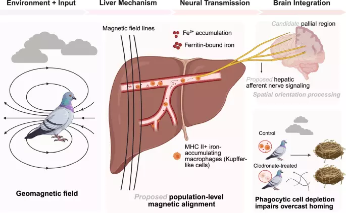
In a recent study published in Science, Lisowski et al. identified superparamagnetic, iron-loaded macrophages in the pigeon liver and showed that depletion of clodronate-sensitive phagocytic cells impaired homing under overcast skies, when solar cues were unavailable.1 Together, these observations point to a light-independent magnetoreceptive system outside the head and broaden current models of magnetoreception beyond receptors based on cryptochrome, magnetite, and ion channels in the eye and beak.2 Magnetic measurements pointed the way (Fig. 1). Vibrating-sample magnetometry revealed a magnetic blocking transition near 12 K in liver and spleen, the signature of superparamagnetic nanoparticles, absent from muscle and beak. The measured signal could not be explained by isolated paramagnetic ferric iron, and low-temperature hysteresis indicated long-range magnetic order: not merely iron in the liver, but a biological nanomagnet.
Prussian blue staining localized ferritin-bound iron to MHC II+ cells in hepatic sinusoids, abundant in liver, sparse in spleen, and absent from brain, eye, muscle, and beak. Identifying the cells required a combination of histology and transcriptomics. Iron and MHC II colocalized, and sorted MHC II+ cells revealed a clear macrophage signature, together with enrichment of Spi-C, a transcription factor central to iron-handling red-pulp macrophages.
Notably, no significant enrichment of dendritic-cell or B-cell signature was detected, an exclusion that matters because MHC II marks all three antigen-presenting lineages. Functionally, the cells phagocytosed dextran with high efficiency, consistent with an active macrophage identity. Together, these data indicate that the iron reservoir is not diffuse but cell-specific: the very macrophages that recycle iron from senescent erythrocytes accumulate ferritin in sufficient quantities to generate measurable magnetism.
Because clodronate can also perturb neutrophils, the authors examined heterophils, the avian equivalent, and found them devoid of iron and of any magnetic blocking, supporting macrophages, rather than granulocytes, as the iron-containing candidate cells. Behavioral experiments then provided the decisive test. Pigeons trained to home over a 19 km route were depleted of clodronate-sensitive phagocytic cells, under conditions favoring hepatic iron-loaded macrophages, and released under fully overcast skies, when solar and polarized-light cues are unavailable.
Control birds returned within ~70 min, whereas treated birds dispersed in random directions and failed to home that day. Crucially, the same birds still homed under sunny conditions, arguing against impaired flight capacity, motivation, or health, and supporting a deficit in magnetic orientation under overcast conditions. The effect was robust across multiple overcast release days over two years.
Anatomically, iron-loaded macrophages were found within ~2 μm of unmyelinated autonomic nerve fibers in the portal triads, a geometry compatible with proposed hepatic afferent nerve signaling. Clodronate treatment disrupted these macrophage–nerve associations while sparing the nerves themselves, supporting the possibility of a relationship between magnetic material and peripheral neural pathways, although direct signal transmission remains to be demonstrated. These findings support macrophage-based magnetoreception as a potential fourth mechanism of magnetic sensing.
Extract — continue reading at the source.