In a recent study published in Science Translational Medicine, Guo et al.1 shed further light on TDP-43 pathology by demonstrating that several transcripts involved in synaptic function and membrane excitability are aberrantly spliced upon depletion of TDP-43. Simultaneous treatment with antisense oligonucleotides targeting these transcripts corrected the splicing abnormalities and restored neuronal function, suggesting that combination therapy could be a promising approach in TDP-43 proteinopathies.1 TDP-43 is an RNA-binding protein involved in key aspects of pre-mRNA splicing, particularly in the repression of cryptic exons that could lead to frameshifts and premature stop codons. While physiologically localized in the nucleus, TDP-43 forms cytoplasmic aggregates in almost all cases of amyotrophic lateral sclerosis (ALS), the most common form of motor neuron disease (MND), and roughly half of the cases of frontotemporal dementia (FTD), leading to a de facto loss of the protein’s function.
TDP-43 pathology is thus regarded as the common downstream event onto which probably multiple pathogenetic mechanisms converge. But what lies even more downstream? Which of the aberrant splicing events affecting the more than 6000 transcripts targeted by TDP-43 actually drive neurodegeneration?
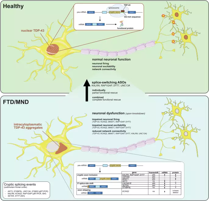
Previous work showed that FTD/MND-associated polymorphisms in UNC13A overlap with TDP-43-binding sites and, upon loss of TDP-43, increase the probability of inclusion of a cryptic exon.2,3 As the protein UNC13A is involved in maturation of synaptic vesicles and neurotransmitter release, these results established a direct mechanistic link between neurodegeneration, TDP-43 pathology and synaptic dysfunction. Building on this work, Guo and coauthors now delve deeper into the downstream landscape of TDP-43 to identify additional targets involved in synaptic function and membrane excitability.1 Leveraging previous transcriptomic datasets from FTD/MND patients, the authors focused on 10 additional transcripts (AKT3, CACNA1E, CEP290, KALRN, KCNQ2, MNAT1, RAP1GAP, SETD5, STXBP5L and SYT7) with roles in synaptic function and/or membrane excitability, besides the previously characterized UNC13A and STMN2, another established TDP-43 target involved in neurite length regulation. All of them could be validated as bona fide TDP-43 targets based on detection of cryptic splicing alterations in these transcripts upon TDP-43 knockdown in human induced neurons and the identification of cryptic splicing regions containing TDP-43-binding sites and/or GU-rich motifs recognized by TDP-43.
Furthermore, cryptic exon inclusion in several of these transcripts (KALRN, KCNQ2, RAP1GAP, SETD5, and SYT7) was detected in neurons from FTD/MND patients showing nuclear depletion of TDP-43, but not in neighboring neurons retaining normal nuclear TDP-43 localization. The authors then showed that knockdown of some of these transcripts, mimicking the reduced expression observed upon TDP-43 depletion, was sufficient to alter neuronal spontaneous firing, neuronal excitability and/or network connectivity. Finally, they went on to develop antisense oligonucleotides (ASOs) to suppress the cryptic exon inclusion in KALRN, RAP1GAP, SYT7 and UNC13A, and observed that combined administration of all ASOs led to complete rescue of the neuronal dysfunction induced by TDP-43 depletion, in terms of spontaneous firing, neuronal excitability and network connectivity.
A graphical scheme summarizing these findings is shown in Fig. 1. Overall, the study further strengthens the link between TDP-43 pathology, cryptic splicing and neuronal dysfunction, while providing a much more nuanced view of the mechanisms underlying this process. Rather than a “one-man show”, in which disruption of a single pathway is solely responsible for neuronal dysfunction, this is likely the sum of multiple contributions from disparate mechanisms, including altered ion channel activity, impaired neurite outgrowth, and defective synaptic vesicle maturation.
Consistent with this, none of the ASOs alone was sufficient to fully restore neuronal function back to control levels. Nevertheless, it is worth pointing out that ASO-mediated splice correction of some transcripts, particularly KALRN, produced substantially greater improvements in electrophysiological parameters (firing rate, number of bursts, network bursts) than correction of the other targets, raising the possibility that specific proteins or pathways may still have a disproportionate impact. This is even more striking when considering that protein levels of KALRN were only modestly rescued.
Extract — continue reading at the source.