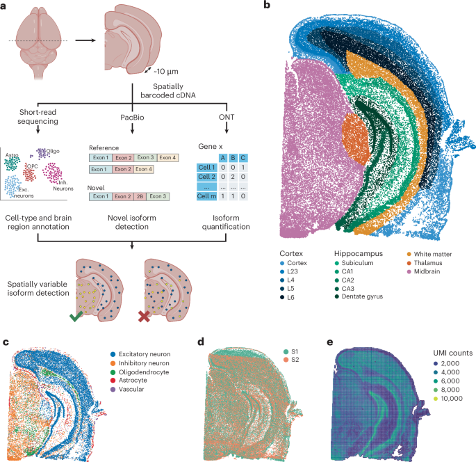
Spatial long-read technologies are increasingly common but usually lack single-cell resolution. This leaves unanswered whether spatially variable isoforms reflect variability within one cell type or differences in region-specific cell-type composition. Here, we developed Spl-ISO-Seq2 (500-nm resolution) and accompanying software, Spl-IsoQuant-2 and Spl-IsoFind, enabling long-read sequencing of >450 million barcodes versus 80,000 previously.
Applying this to the adult mouse brain, we compared differential isoform abundance between known regions and spatial isoform patterns independent of predefined regions. Both identified overlapping hits, for example, Rps24 in oligodendrocytes. For known Snap25 spatial isoform variation, we show that it occurs in excitatory neurons.
The region-agnostic approach also uncovered patterns missed by region-based comparisons, for example, for Ighm. Notably, many spatial isoform signals are not driven by cell-type composition alone. Finally, our software is applicable to many spatial and single-cell protocols, demonstrating reproducibility between platforms (for example, Visium HD/Stereo-seq).
Overall, our experimental/analytical methods enable a submicron-resolution-isoform view and open avenues for spatial isoform disease research. A fundamental question in spatial isoform biology is whether a cell’s spatial position influences its isoform expression. This question can be confounded by different cell types dominating distinct areas in the brain.
Therefore, single-cell-resolution spatial isoform data are essential for disentangling spatially distributed isoforms from cell-type-derived ones. Single-cell and single-nucleus long-read sequencing have enabled profiling isoforms in distinct cell types1,2,3,4,5. Short-read sequencing-based approaches have also described cell-type-specific splice junctions6,7,8, revealing exons that are used in specific cell types or altered in evolution and/or disease5,9,10; however, a major shortcoming of these methods is that they lose the spatial location of cells, preventing the study of how isoforms may be spatially regulated at the cell-type-specific level.
Simultaneously, spatial gene-expression profiling has moved the field forward by localizing cell types and gene expression across tissue sections11,12,13,14,15,16,17,18. Based on 10x Genomics’ Visium technology, we and our colleagues simultaneously engineered spatial isoform sequencing, revealing many isoform switches that correlated tightly with brain structures, and other isoforms (for example, of Snap25) that showed isoform changes within a region4,19; however, Visium’s 55-μm spot size exceeds the average cell diameter in the mouse brain, and as such, results in ‘pseudo-bulk’ measurements that likely represent multiple cells and thus potentially multiple cell types. By adapting Slide-SeqV2 (ref. 12), we further developed spatial isoform sequencing (Spl-ISO-Seq) with 10-μm resolution and corresponding software Spl-IsoQuant20.
Extract — continue reading at the source.