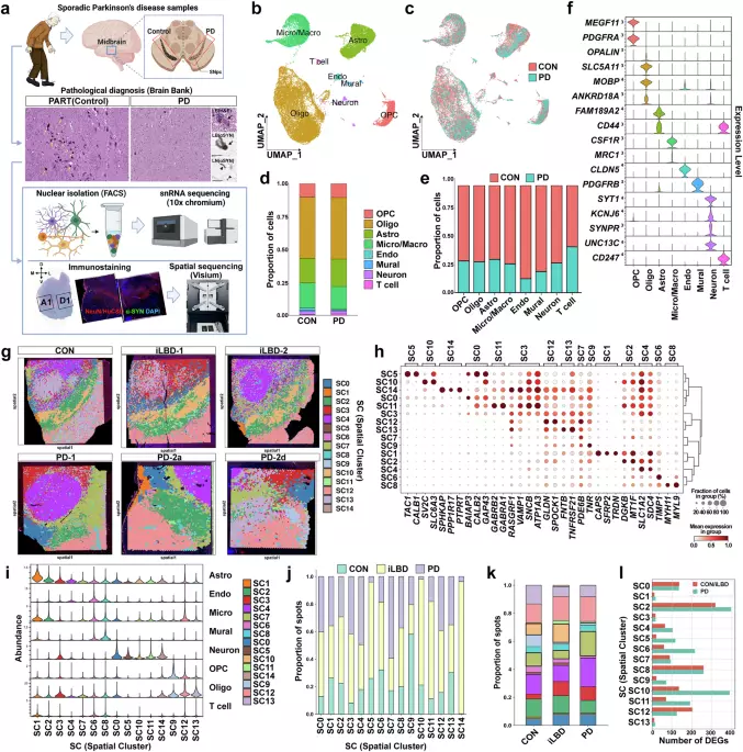
Parkinson’s disease (PD) is clinically and biologically heterogeneous, and ethnic differences may influence its pathogenesis. However, single-nucleus transcriptomic data from East Asian populations remain limited. Here, we present a spatially resolved single-nucleus transcriptomic atlas of the human substantia nigra (SN) across the PD continuum, control, incidental Lewy body disease (iLBD), and advanced PD, in pathologically confirmed Korean individuals.
Our integrative analysis revealed early α-synuclein aggregation in the ventrolateral SN, subtype-selective vulnerability of dopaminergic neurons, and relative preservation of the dorsomedial SN and ventral tegmental area. Trajectory and gene regulatory network analyses of astrocytes and microglia revealed spatiotemporally distinct, disease-associated glial responses, each driven by divergent molecular programs. We also identified two transcriptionally distinct vascular spatial clusters that exhibited differential vulnerability and remodeling.
Spatial ligand–receptor analysis of α-synuclein-positive regions revealed intercellular signaling events associated with early pathology, including extracellular matrix remodeling and synaptic adhesion, which were prominent in the iLBD but largely diminished in advanced PD. Integration with PD genome-wide association studies using MAGMA demonstrated differential coupling between genetic risk, spatially resolved cell-state programs, and pseudotime-inferred microglial dynamics across ancestries. This study provides the first high-resolution, spatially mapped single-nucleus transcriptomic resource from an East Asian PD cohort, illuminating early perturbations in the midbrain intercellular and microenvironment that may underlie selective dopaminergic vulnerability and guide the development of ancestry-informed therapeutic strategies.
PD is the second most common neurodegenerative disorder and is characterized by the abnormal accumulation of alpha-synuclein (α-Syn), which leads to Lewy body formation and the progressive loss of dopaminergic neurons in the substantia nigra (SN) of the midbrain. Degeneration of these neurons disrupts basal ganglia circuitry and ultimately produces the characteristic motor symptoms of PD. As with other neurodegenerative diseases, early detection of PD remains challenging because clinical symptoms typically emerge only after substantial dopaminergic neuron loss has already occurred.
Although clinical diagnosis relies on observable motor symptoms such as bradykinesia, rigidity, tremor, and postural instability, a definitive diagnosis still depends on neuropathological confirmation from postmortem brain tissue. In addition to these motor manifestations, PD is frequently accompanied by a broad range of non-motor symptoms, including cognitive impairment, sleep disturbances, and autonomic dysfunction. These features reflect the widespread impact of PD pathology beyond the nigrostriatal pathway and highlight the complex and heterogeneous nature of the disease.
Importantly, neuropathological studies indicate that neuronal vulnerability in PD is not uniform across the SN but instead follows a spatially selective pattern, suggesting that local cellular environments and regional molecular programs may influence disease susceptibility. Understanding the molecular architecture of vulnerable regions within the human midbrain is therefore essential for clarifying the mechanisms that drive selective dopaminergic degeneration and cellular pathways that may contribute to disease initiation and progression. Genetic studies, including pedigree analysis, whole-exome sequencing, and genome-wide association studies (GWAS), have yielded valuable insights into PD-associated genetic risk factors, some of which have been identified as causal and are currently being explored as therapeutic targets.1,2 Variants in genes such as SNCA, LRRK2, GBA1, and PARK2 have been implicated in PD pathogenesis, and several targeted therapeutic strategies are under active investigation.
Extract — continue reading at the source.