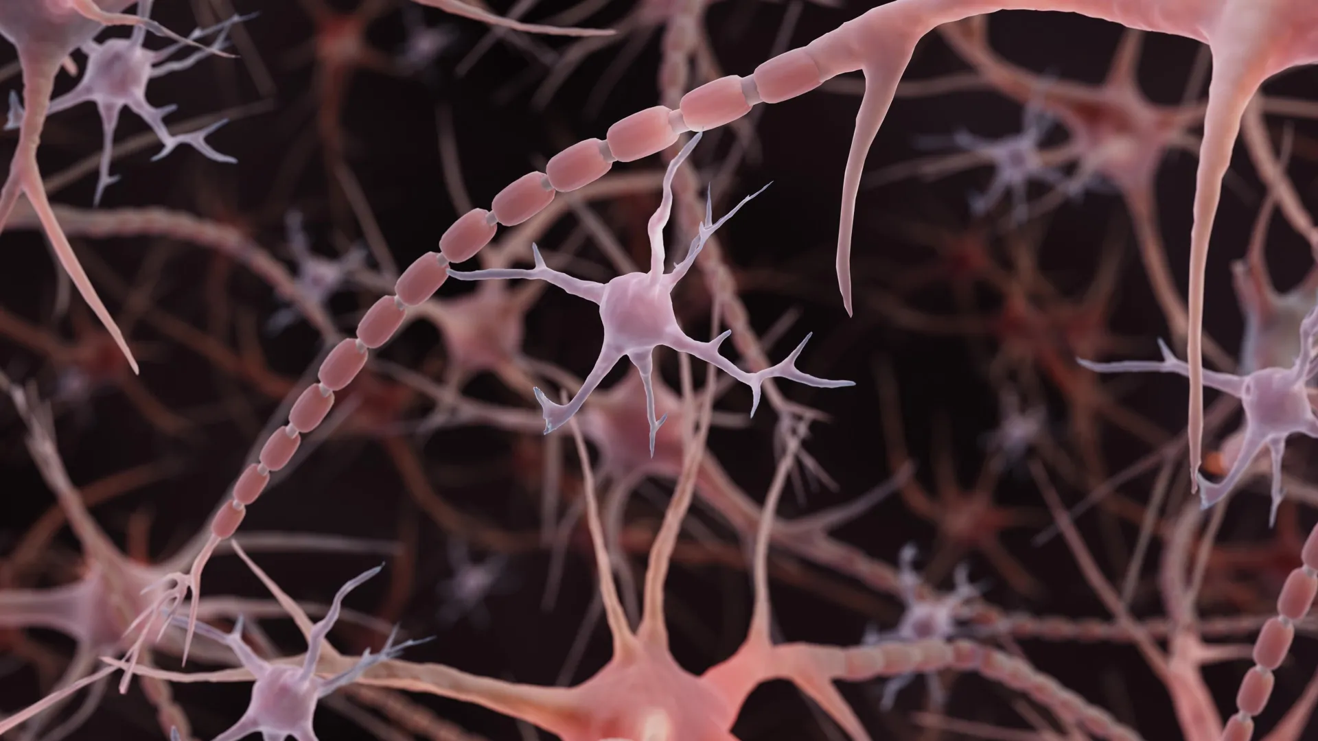
The brain's microscopic wiring may need a different kind of textbook illustration. Instead of the smooth, narrow tubes commonly drawn extending from nerve cells, some axons resemble strings of tiny pearls. These repeating bulges are not simply decorative details.
Their shape appears to influence how electrical messages travel. Researchers at Johns Hopkins Medicine brought this unexpected architecture into focus in a study published online in Nature Neuroscience on December 2, 2024. Working with mouse neurons, they found evidence that a familiar picture of brain cell anatomy may overlook an important feature of the cells' signal-carrying extensions.
Related research has since documented pearled axons in human brain tissue, extending the observations beyond mice. Together, the findings raise questions about how the physical structure of the brain's wiring helps regulate communication. "Understanding the structure of axons is important for understanding brain cell signaling," said Shigeki Watanabe, Ph.D., an associate professor of cell biology and neuroscience at the Johns Hopkins University School of Medicine, in the university's 2024 announcement.
"Axons are the cables that connect our brain tissue, enabling learning, memory and other functions." Axons are the long extensions that allow neurons to transmit electrical signals to other cells. The conventional illustration shows a relatively uniform tube interrupted by occasional bulges (synaptic varicosities that hold globs of neurotransmitters, which enable signaling to other brain cells). Beaded axons themselves are not an unfamiliar sight.
Scientists have long observed pronounced swellings in dying neurons and in neurodegenerative conditions, including Parkinson's disease. In those settings, the beading can accompany damage to the cell membrane and the internal framework that helps maintain the axon's structure. What stood out in the Johns Hopkins study was the presence of much smaller, repeating swellings in axons examined under conditions intended to preserve their normal structure.
The researchers called these regions "non-synaptic varicosities." Unlike the familiar bulges associated with communication sites, these swellings were not synapses. The distinction matters: the study did not suggest that every beaded axon is damaged. Instead, it pointed to a form of nanoscale pearling that may be part of the normal architecture of the axons studied.
Extract — continue reading at the source.


